Microscopy platforms
Four complementary imaging systems covering confocal, widefield, and live-cell applications. All require advance reservation and completed training.
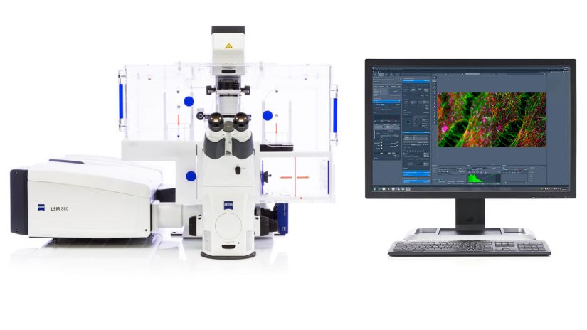
7 laser lines. 32-channel GaAsP Airyscan detector, 32-channel QUASAR spectral detector. Fixed and live-cell imaging.
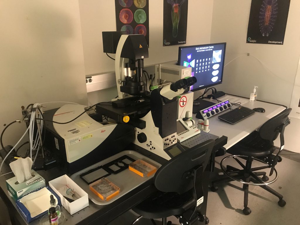
White light laser (470–670 nm), resonant scanner, HyD detectors, Lightning deconvolution. NIH-funded via S10 OD016240.
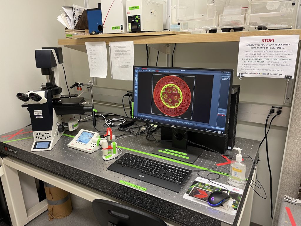
Motorized stage for mosaic tiling, DIC optics, full fluorescence filter sets. Ideal for large-area and multi-point experiments.
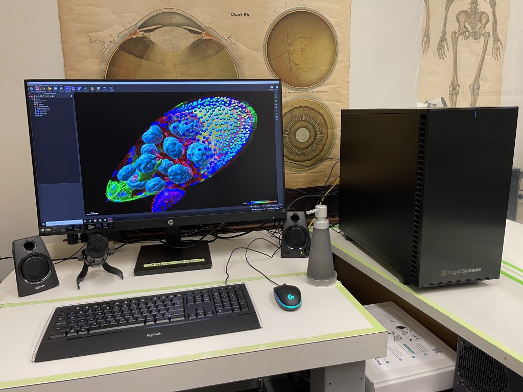
Full-license 3D/4D suite on AMD Threadripper — surface rendering, spot detection, filament tracing, cell analysis. Free for UW students.
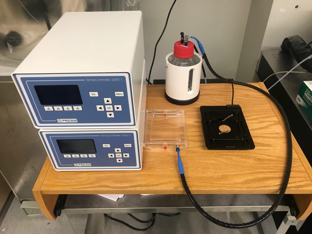
Pecon stage-top incubator and Warner 35 mm dish incubator. Temperature, CO₂, and humidity control for live-cell experiments.
Zeiss LSM 880 — detailed specs
- 405 nm, 440 nm diode laser
- 458 nm, 488 nm, 514 nm Argon laser
- 561 nm DPSS laser
- 633 nm HeNe laser
- 32-channel GaAsP spectral detector for simultaneous spectral imaging
- 2× PMT detectors for standard fluorescence channels
- Airyscan detector — 1.7× resolution improvement over diffraction limit
- Bi-directional scanning up to 8 fps at 512×512
- Transmitted light detector for DIC/brightfield
- ~140 nm lateral resolution, ~400 nm axial resolution
- Improved signal-to-noise ratio compared to conventional confocal
- Compatible with all laser lines and standard fluorophores
- ZEN Black — acquisition, processing, and analysis
- Spectral unmixing for multi-fluorophore experiments
- FRAP, FRET, and time-series modules
- 3D rendering and orthogonal projection
- 10×/0.45 air — large field of view, tissue sections
- 20×/0.80 air — standard fluorescence
- 40×/1.30 oil — high-resolution subcellular
- 63×/1.40 oil — fine subcellular detail, Airyscan super-resolution
- 100×/1.45 oil — maximum resolution, single-molecule imaging